Central Instrumentation Facility Unit (CIFU)
The Central Instrumentation Facility Unit came into existence at CSIR-NIIST on the 1st of June 2023. This facility was created to provide a well streamlined, professionally managed analytical support through a single window concept to the ongoing in-house research projects, to support the analytical requirement of research scholars pursuing their PhD program at CSIR-NIIST and to external clientele comprising of Industries, MSME, Start-ups, Small Scale Industries and students from universities and colleges pan India.
CIFU functions as a platform dealing with the queries for availing analytical services, processing of samples for analysis, handling customer requirements and concerns, management and maintenance of the sophisticated analytical facilities.
CSIR-NIIST has more than 200 plus analytical facilities operated through dedicated operators/technicians backed by our scientist support.
We have the integrated NABL accreditation for Dioxins link PoP Testing analysis in the environment, Food and Feed samples etc.
We also undertake the biodegradability testing and analysis of biodegradable items as per the ISO 14855 and ISO 14985 test methods.
Procedure for sample submission:
The external samples may be submitted to the following address of CIFU through SPEED POST only.
The Head
Central Instrumentation Facility Unit
CSIR-NIIST, Industrial Estate PO
Thiruvananthapuram 695019
Kerala State
Before submission of the sample please ensure that your analytical requirement is feasible at CSIR-NIIST by sending details of your analytical requirement to the email id: chandra@niist.res.in
While sending your email, please ensure that the details of samples (nature of the sample, type of solvent to be used-wherever applicable, column preferred in case chromatography services are availed etc) need to be clearly mentioned.
Please ensure that the exact billing address with GST details (if applicable) is mentioned in the covering letter to issue the invoice against the payment made.
The external samples will be taken in for analysis on a first to come first to serve basis.
Users are requested to pay the charges along with applicable taxes in advance (before the analysis of the sample)
The client/customer/student seeking the services through the Central Instrumentation Facility Unit shall indemnify and hold CSIR/CSIR-NIIST harmless against any and all damages, liability, law suits, demands, which may arise by the use of data, information, interpretation of analysis results provided by CSIR-NIIST on the sample submitted.
People

Shri. Chandrakanth C.K.
Senior Principal Scientist & Head- 9061082354
- chandra[at]niist[dot]res[dot]in

Shri. Kiran J.S.
Technical Officer- 8547489600
- kiranjs[at]niist[dot]res[dot]in

Dr. Guguloth Venkanna
Technical Assistant- 9676347020
- gvenkanna[at]niist[dot]res[dot]in
| SI.NO | Instrument Name | Instrument principle | Inventor | Analysis | Instrumentation |
|---|---|---|---|---|---|
| 1 | Flame Photometer | The principle of flame photometer is based on the measurement of the emitted light intensity when a metal is introduced into the flame. The wavelength of the colour gives information about the element and the colour of the flame gives information about the amount of the element present in the sample. |
Champion, Pellet, and Grenier
|
Flame photometer is an analytical instrument used in clinical laboratories for determining of sodium, potassium, lithium and calcium ions in body fluids |
 |
| 2 | ICP | Inductively Coupled Plasma Optical Emission spectroscopy (ICP-OES) is an analytical technique used to determine how much of certain elements are in a sample. The ICP-OES principle uses the fact that atoms and ions can absorb energy to move electrons from the ground state to an excited state. |
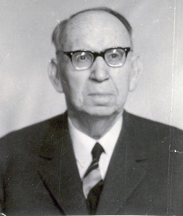
Eugen Badara
|
ICP (Inductively Coupled Plasma) Spectroscopy is an analytical technique used to measure and identify elements within a sample matrix based on the ionization of the elements within the sample. |
 |
| SI.NO | Instrument Name | Instrument principle | Inventor | Analysis | Instrumentation |
|---|---|---|---|---|---|
| 1 | Amino Acid Analyzer | Amino acid analysis is a technique based on ion exchange liquid chromatography, used in a wide range of application areas to provide qualitative and quantitative compositional analysis. The basic principle of operation is the continuous flow chromatography procedure developed by Spackman, Moore and Stein in 1958. |
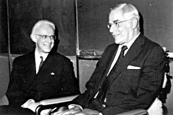
Spackman, Moore and Stein
|
Amino acid analyser is an analytical instrument to analyse the amino acids in body fluids such as urine, serum, and blood and in food samples. The measurement of amino acids is an essential element of physiological and nutritional studies and for monitoring the growth of cells in cultures |
 |
| 2 | DNA Sequencing-750-850bp | This method is based on the principle that single-stranded DNA molecules that differ in length by just a single nucleotide can be separated from one another using polyacrylamide gel electrophoresis, described earlier. One dideoxynucleotide, either ddG, ddA, ddC, or ddT. |
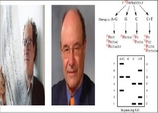
Allan Maxam and Walter Gilbert
|
Sequence analysis is a term that comprehensively represents computational analysis of a DNA, RNA or peptide sequence, to extract knowledge about its properties, biological function, structure and evolution. |
 |
| SI.NO | Instrument Name | Instrument principle | Inventor | Analysis | Instrumentation |
|---|---|---|---|---|---|
| 1 | X-Ray Radiography | It is based on the principle that radiation is absorbed and scattered as it passes through an object. If there are variations in thickness or density (e.g. due to defects) in an object, more or less radiation passes through and affects the film exposure. Flaws show up on the film, usually as dark areas. |
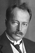
Max von Laue
|
X-Ray diffraction analysis (XRD) is a non-destructive technique that provides detailed information about the crystallographic structure, chemical composition, and physical properties of a material |
 |
| 2 | AAS | AAS is an analytical technique used to determine how much of certain elements are in a sample. It uses the principle that atoms (and ions) can absorb light at a specific, unique wavelength. When this specific wavelength of light is provided, the energy (light) is absorbed by the atom. |
Alan Walsh
|
AAS is an analytical technique used to determine how much of certain elements are in a sample. It uses the principle that atoms (and ions) can absorb light at a specific, unique wavelength. When this specific wavelength of light is provided, the energy (light) is absorbed by the atom. |
 |
| SI.NO | Instrument Name | Instrument principle | Inventor | Analysis | Instrumentation |
|---|---|---|---|---|---|
| 1 | GC-MS | A mixture will separate into individual substances when heated. The heated gases are carried through a column with an inert gas (such as helium). As the separated substances emerge from the column opening, they flow into the MS. |
Robert Emmet Finnigan
|
Analysis begins with the gas chromatograph, where the sample is effectively vaporized into the gas phase and separated into its various components using a capillary column coated with a stationary (liquid or solid) phase. |
 |
| 2 | Kjeldahl Apparatus | Strong acid helps in the digestion of food so that it releases nitrogen which can be determined by a suitable titration technique. |
Johann Kjeldahl
|
Used to routinely measure the crude protein content of foods. |
 |
| SI.NO | Instrument Name | Instrument principle | Inventor | Analysis | Instrumentation |
|---|---|---|---|---|---|
| 1 | AFM | The underlying principle of AFM is that this nanoscale tip is attached to a small cantilever which forms a spring. As the tip contacts the surface, the cantilever bends, and the bending is detected using a laser diode and a split photodetector. This bending is indicative of the tip-sample interaction force. |
Binnig, Quate, and Gerber
|
Atomic Force Microscopy (AFM) analysis provides images with near-atomic resolution for measuring surface topography. AFM is also referred to as Scanning probe microscopy. Atomic Force Microscopy is capable of quantifying surface roughness of samples down to the angstrom-scale. |
 |
| 2 | HRTEM | An electron source usually named as the “Gun” produces a stream of electrons which is accelerated towards the specimen using a positive electrical potential. This stream is then focused using metal apertures and magnetic lenses called “condenser lenses” into a thin, focused, monochromatic beam. |
Ernst Ruska and Max Knoll
|
HRTEM is an imaging mode of the TEM that allows the imaging of the crystallographic structure of a sample at an atomic scale. |
 |


